This section is a review of basic spine anatomy. It covers the bones, ligaments, muscles and other structures that make up and support the spine. The information is applicable for both back anatomy and neck anatomy. For more information on how the spine works please read the section on basic spine biomechanics.
What are the different sections of the spine?
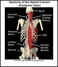
The spine is one of the most complex parts of the body. The spine can be divided into five sections: the cervical section (the neck), the thoracic section (the upper back), the lumbar section (the lower back), the sacrum (part of the pelvis) and the coccyx (the tailbone).
Anatomy of a Typical Vertebra
Each section of the spine contains a number of “back bones” called vertebrae. A typical vertebra has a “body”, two laminae, two transverse processes, a spinous process and four facets (see picture). The body is the main part of the vertebrae and it is designed to carry weight. The laminae form the bony tunnel that allows the spinal cord to pass down the spine. The paired transverse processes and the spinous process are bony extensions of the spine and act as attachment sites for muscles and ligaments. The facets are paired, flat areas of the vertebrae that form joints (facet joints) with the facets of the vertebrae above and below.
Vertebrae in the cervical, thoracic and lumbar sections of the spine are separated by a structure called the “intervertebral disc”. The discs act as spacers between the vertebrae and they also function as shock absorbers. Each disc consists of a spongy center called the “nucleus pulposis”, and a tough outer layer called the “annulus fibrosis”.
How many vertebrae are in the different sections of the spine?
Although the vertebrae in the spine are similar, each section of the spine has vertebrae with unique structural features. The smallest moving vertebrae in the spine are located in the cervical section. There are 7 cervical vertebrae. These vertebrae are designed for motion rather than weight-bearing (the first cervical vertebrae is not a typical vertebrae in that it does not have a vertebral body or a disk above it). In the thoracic section of the spine there are 12 vertebrae. These vertebrae are larger than the cervical vertebrae. Each thoracic vertebra has attachment sites for the left and right ribs. In the lumbar section of the spine there are 5 vertebrae. These vertebrae are the largest in the spine and are primarily designed for supporting the weight of the body.
The sacrum is a single bone that also forms part of the pelvis. This triangular shaped bone is actually 5 fused vertebrae. It plays a role in transferring the weight of the spine and upper body to the hips. The coccyx is also a single bone that consists of 4 small fused vertebrae. It attaches to the bottom of the sacrum.
Ligaments are like strong ropes that help connect bones and provide stability to joints. Six major ligament groups run down the spine. These ligaments attach to each vertebra and form a strong support system for the spinal column.
What is the function of the spine?
The complex anatomy of the spine allows it to perform various functions. The spine supports the weight of the body, protects the spinal cord and it allows the body to move by providing attachment sites for muscles. The muscles located closest to the vertebrae help to provide stability to the spine. The muscles located further from the vertebrae play a role in movement of the spine.
Finally, the spine protects the spinal cord and allows the spinal nerves to exit the spinal column. The spinal nerves exit through openings in the spinal column called the intervertebral foramen. There is a left and right intervertebral foramen between vertebrae. These openings allow the nerves to pass through them and transfer information between the spinal cord and the specific tissues and organs they supply.
A recommended book for learning spine anatomy:
Moore’s Clinically Oriented Anatomy
Renowned for its comprehensive coverage and engaging, storytelling approach, the bestselling Moore’s Clinically Oriented Anatomy, 9th Edition, guides students from initial anatomy and foundational science courses through clinical training and practice. A popular resource for a variety of programs, this proven text serves as a complete reference, emphasizing anatomy that is important in physical diagnosis for primary care, interpretation of diagnostic imaging, and understanding the anatomical basis of emergency medicine and general surgery.